

Location.com logo
Comprehensive Neurosurgery | Lewisville, TX
Brand Certified

Comprehensive Neurosurgery

4.4
(65 reviews)

Business Details

1700 FM 544, Lewisville, TX
75056, United States
(972) 394-4600
https://comprehensiveneurosurgery.com/

About

Neurosurgery
Comprehensive Neurosurgery takes pride in the concern for our patients’ quality of life. Prevention education, diagnosis, surgical treatment, and rehabilitation are examples of the direct approaches to quality life improvement. Comprehensive Neurosurgery strives for effective patient-centered improvements with compassion and integrity being our number one priority. Having one goal in mind and unity, we will be exactly what you’re looking for in your neurosurgery needs.

Location

Comprehensive Neurosurgery
1700 FM 544, Lewisville, TX
75056, United States

Hours

Monday8:00 AM - 5:00 PM
Tuesday8:00 AM - 5:00 PM
Wednesday8:00 AM - 5:00 PM
Thursday8:00 AM - 5:00 PM
Friday8:00 AM - 5:00 PM
SaturdayClosed
SundayClosed

Products & Services

1 list · 9 items

Comprehensive Neurosurgery Procedures and Services

9 items

Back Pain
Back Pain

Back Pain Lower Back Pain Lower back pain is the most common cause for a work-related disability and one of the main reasons for people staying home from work. The lower back is one of the most important parts of the body as it holds most of our body weight when we stand and is involved in movement when we bend or twist at the waist. Because of its pivotal role and frequent use, the lower back is susceptible to injury and chronic pain. Lower back pain is especially common in older adults who may have decreased bone strength and muscle elasticity. The spongy cartilage pads, called intervertebral discs, that allow for flexibility may wear away and weaken in an elderly person. Causes Of Lower Back Pain Lower back pain can be a result of many different reasons including the following: -Muscle sprain, strain or spasm -Irritated nerve -Bacterial or viral infection -Obesity -Smoking -Poor posture -Sleep position -Pain may radiate down the legs as well. Conditions Associated With Lower Back Pain There are some conditions that cause lower back pain. They include: -Bulging disc -Sciatica -Degeneration of the spine -Spinal Stenosis -Osteosporosis -Spondylolisthesis -Fibromyalgia -Spinal irregularity -Osteoarthritis -Neuropathy -Spinal fracture -Deformity of the spine -Tumor -Diagnosis Of Lower Back Pain While lower back pain is a common condition, it may also be an indication of another condition which may be far more serious. After obtaining a medical history, the physician may conduct the following tests to confirm diagnosis and determine treatment: -X-ray imaging -Discography -CT scan -MRI -EMG -Bone scan -Ultrasound -Treatment Of Lower Back Pain While lower back pain is not usually a serious condition, it can be very painful and debilitating. Treatment for this pain is usually simple and can include: -Medication -Ice -Heat -Rest -Exercise -Chiropractic spinal adjustment -Acupuncture -Biofeedback -Interventional therapy -Traction -Ultrasound -TENS -Surgery For those people who suffer from lower back pain as a result of either improper body mechanics or a non-traumatic event, there are some ways to prevent lower back pain. Some of them include: -Stretching before exercising -Ensuring posture is correct -Good lumbar support when in a seated position -Wearing heels that are low and comfortable -Sleeping on your side to prevent a curvature of the spine -Not lifting objects that are too heavy -Maintaining a healthy weight -Maintaining a healthy lifestyle -Quitting smoking -Back Pain Prevention At some point in their lives, the great majority of adults will suffer from significant low back pain, usually from an injury at work, at home or at play. Orthopedists, chiropractors, physical therapists, coaches and trainers all have helpful advice regarding back pain prevention. By following their directives, people can minimize the possibility of back injury, and keep themselves healthier in the process. Lifestyle Changes For Back Pain Prevention Maintaining a healthy lifestyle can go a long way in preventing acute injuries or chronic back pain. Taking the following steps can help maintain strength, balance and flexibility, and promote rapid healing if an injury occurs: -Exercise regularly for strength and conditioning -Eat a nutritious diet -Maintain a healthy weight -Do not smoke -Do not drink alcohol to excess -Do not abuse drugs Following these directives helps to keep muscles well-toned and bones at optimal density. It also prevents excess weight from putting undue strain on muscles and spinal discs. It is essential to also pay attention to emotional health; depression, anxiety and psychic stress can make people prone to injury, slow to recover and overly sensitive to pain. Posture And Ergonomics For Back Pain Prevention Learning proper posture and using ergonomic furniture/equipment are also important in preventing back pain. It is beneficial to learn appropriate ways to stand, sit, bend, lift and turn, so as not to compromise spinal health. Using assistive devices when necessary for reaching or lifting is recommended, preventing physical stress that can result in injury or long-term damage. Becoming aware of the initial signs of pain can help trace its origins. Noticing that pain occurs after sitting in a particular chair, wearing particular shoes, performing a particular task, or reaching for something in a certain way, can be of diagnostic significance. It should also be a warning that a pattern of movement or behavior should be changed. In some cases, replacing furniture or equipment with more ergonomically designed pieces may be necessary. It may also be possible to find a more convenient, less expensive solution to a problem by supplementing a chair or car seat with a supportive back cushion, or getting orthotics for one’s shoes.
Degenerative Disk

Degenerative Disc Degenerated discs are a common back problem. The spinal discs, which are soft, gelatinous cushions that separate the vertebrae, wear down during the aging process. Because the discs function as between-the-bones shock absorbers, allowing the spine to bend and twist, this deterioration can result in serious back pain. As discs are damaged or wear away, the amount of space between the vertebrae gets smaller. As the space narrows, joints are placed under greater stress, resulting in further degeneration. Risk Factors For Degenerated Discs Although discs degenerate to a degree from normal aging, there are risk factors that increase the likelihood of symptomatic disc degeneration. They include the following: -Repeated heavy lifting -Obesity -Smoking -Bone spurs -An acute injury to the back also increases the potential for disc degeneration. Symptoms Of Degenerated Discs Compression of the spinal nerves can cause symptoms that include tingling/numbness, muscle weakness, loss of balance/ coordination, and pain and stiffness. Back pain is often worse when sitting, lifting, bending or twisting, and walking or lying down typically helps alleviate the pain. Diagnosis Of Degenerated Discs In order to diagnose a degenerated disc, a physician takes a medical history, and performs a comprehensive physical examination that includes checking for numbness or weakness, and testing reflexes to determine whether any muscles have atrophied. Several imaging examinations may also be administered; they include X-rays, to assess spine alignment; MRI scans, to observe any compression of the spinal cord; and CT scans, to evaluate the size of the spinal canal and visualize bone spurs. A myelogram, which uses contrast dye for improved visualization during X-rays or CT scans, may also be administered. Complications Of Degenerated Discs Although a disc can degenerate anywhere along the 26 vertebrae of the spine, it happens most frequently in the lumbar region (lower back) or the cervical region (neck). In addition to pain, degenerated discs can result in complications that include the following: -Osteoarthritis -Herniation of a disc -Spinal stenosis (narrowing of the spinal canal) -Bone spurs -Any of these complications can worsen the patient’s condition. Treatment Of Degenerated Discs In many cases, nonsurgical options provide a patient with relief from a degenerated disc. Bed rest and anti-inflammatory medications can help decrease swelling and pain. If the affected disc is in the cervical region, a soft collar can limit range of motion, and alleviate pressure on nerve roots. Physical therapy may be recommended to stretch and strengthen the muscles of the abdomen, back and, possibly, the neck. Epidural injection of corticosteroids can help to reduce swelling, and greatly relieve pain in the affected area. A relatively new form of minimally invasive treatment is intradiscal electrothermal therapy, during which a catheter with a heating coil is inserted through a needle into the affected disc. The heat strengthens the collagen fibers that hold the disc together, strengthening tissue while destroying excess nerve fibers. Intradiscal electrothermal therapy is often successful in decreasing damage and pain. If a patient’s back pain does not respond to these conservative treatments, surgical intervention may be required. If a disc has become herniated and is pressing on a nerve, a discectomy may be recommended to remove the disc entirely. The removed disc may be replaced by a synthetic disc that can restore shock absorption and improve mobility between vertebrae. In more severe cases, spinal-fusion surgery, which involves removing the disc and fusing the nearby vertebrae to increase stability in the spine, may be performed.

Spinal Stenosis
Spinal Stenosis

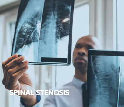
Spinal Stenosis Spinal stenosis is the narrowing in one or more areas of the spinal canal as a result of injury or deterioration of the discs, joints or bones of the spine. Most cases of spinal stenosis develop as a result of the degenerative changes that occur during aging. Osteoarthritis is the main cause of spinal stenosis since this condition causes deterioration of cartilage in the area that leads to the bones rubbing against each other. As bones make repeated abnormal contact, bone spurs form, narrowing the spinal canal. Other causes of spinal stenosis are traumatic injury, herniated disc, ligament thickening and, in rare cases, spinal tumors, any of which can damage the alignment of the vertebrae. A subtype of spinal stenosis is foraminal stenosis. This condition is caused by a narrowing of the foramen, the opening within each of the spinal bones that allows nerve roots to pass through. Symptoms Of Spinal Stenosis Patients with spinal stenosis may experience a number of troubling symptoms. These may include: Pain in the back, neck, shoulders or extremities Muscle cramping Loss of sensation in affected areas Loss of balance Bladder or bowel dysfunction The loss of bladder or bowel control is a rare, but particularly distressing symptom, for which surgery is most often necessary. Diagnosis Of Spinal Stenosis In order to diagnose spinal stenosis, a medical history and a physical examination are always necessary. The condition is often difficult to diagnose, not only because its symptoms may resemble the symptoms of other conditions, but because they may only occur intermittently. A diagnosis of spinal stenosis is usually achieved only after ruling out other disease conditions. Typically, imaging exams such as spinal X-rays, MRI, CT or bone scans are administered to definitively diagnose the condition and to pinpoint the spinal region affected. An electromyography (EMG) may also be administered to measure electrical impulses in the affected skeletal muscles. Treatment Of Spinal Stenosis Most cases of spinal stenosis can be effectively treated through conservative methods such as physical therapy, nonsteroidal anti-inflammatory drugs (NSAIDs), rest and a supportive back brace or cervical collar. In some situations, corticosteroid injections may provide relief. For more severe cases, surgical procedures such as a decompressive laminectomy, laminotomy or spinal fusion may be required to relieve pressure on the nerves. When the patient is diagnosed with foraminal stenosis, a surgical procedure known as a foraminotomy may be performed. This operation widens the area where the spinal nerve roots project from the spine.

Brain Tumor
Brain Tumor

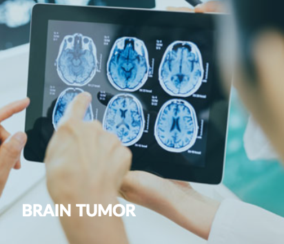
Brain Tumor Brain tumors can be classified in two ways. Primary brain tumors originate in the brain or the spinal canal, whereas secondary tumors spread from other areas of the body to the brain or the spinal canal. Approximately 25 percent of brain tumors are secondary, and all secondary brain tumors are malignant. The majority of secondary brain tumors are caused by lung cancer that has metastasized. Primary brain tumors, however, may be benign or malignant. They develop from different types of brain tissue, and are often named after the areas in which they originate. Primary benign tumors usually grow very slowly; primary malignant tumors usually grow rapidly and are life-threatening. Although a primary malignant brain tumor is sometimes referred to as a “brain cancer,” unlike most types of cancer, it rarely spreads to other parts of the body. It can, however, spread within the brain and spinal cord. Risk Factors For Primary Brain Cancer The specific cause of primary brain cancer, and why it develops in some people rather than others, is unknown. It is believed, however, that certain factors may increase the risk of developing it. Some of these risk factors include: -Family history of brain cancer -Cigarette smoking -Exposure to ionizing radiation -HIV infection -Exposure to environmental toxins Ionizing radiation is produced by cancer-treating radiotherapy and atomic bombs. There is no proven link between brain cancer and radiation from microwaves or cell phones. Symptoms Of Brain Tumors The symptoms of both a benign and malignant brain tumor are usually caused by the swelling and inflammation caused by the growing tumor, and by its pressing on other parts of the brain, which prevents the brain from functioning as it should. However, brain tumors may also be asymptomatic. Common symptoms of both benign and malignant brain tumors include: -Headache -Weakness -Seizures -Difficulty walking -Clumsiness Because they tend to have defined margins, and be rooted less deeply than malignant brain tumors, benign brain tumors are easier to remove surgically. Types Of Primary Brain Tumors Accounting for approximately half of all cases, gliomas are the most common primary brain tumors. They also account for 80 percent of malignant primary brain tumors. Other types include: -Meningiomas -Medulloblastomas -Vestibular schwannomas -Pineal gland and pituitary gland tumors -Primary central-nervous-system lymphomas The most common type of primary malignant brain tumor in children is medulloblastoma. Most cases occur in boys, and before the age of 10. Diagnosis Of Brain Tumors The first step in diagnosing brain tumors involves getting a thorough neurological exam, in which, among other things, eye movement, the optic nerve, and balance and coordination are checked. If results of a neurological exam indicate a possible tumor, the following tests may be performed: -CT scan -MRI scan -PET scan -X-ray -Various laboratory tests If the tests above indicate the presence of a brain tumor, either a surgical biopsy or a stereotactic biopsy is performed; both remove a sample of the tumor for microscopic examination. Treatment For Brain Tumors Treatment for brain tumors varies based on a number of factors: -Tumor location -Tumor size -Tumor type -Patient’s age and medical history Treatments for both malignant and benign brain tumors include: -Surgery -Chemotherapy -Radiation therapy -Stereotactic radiosurgery -Steroids Surgical removal is the preferred treatment for a brain tumor, unless the tumor is in an area of the brain that makes surgical removal impossible. Side Effects Of Treatment For Brain Tumors Depending upon the type of treatment used, side effects may include: -Headaches -Nausea -Vomiting -Fatigue -Hair loss -Blood clots -Edema Even with aggressive treatment, the long-term survival rate (5 or more years) for primary brain cancer is only 10 percent. A patient with secondary brain cancer is more apt to die from the original cancer than from brain cancer.

Spinal Tumor

Spinal Tumor Spinal tumors are abnormal growths of tissue found in and around the spinal cord and its surrounding structures. Most spinal tumors are found in the center of the back. Tumors that originate in the spine are called “primary”; tumors that originate elsewhere are called “secondary.” Breast, lung, prostate and renal cancers are the most common types of cancer to spread to the spine. Primary spinal tumors, which can be benign or malignant, are rare. Types Of Spinal Tumors There are three types of spinal tumors, all of which can cause back pain. Intradural-Extramedullary Tumors Intradural-extramedullary tumors grow within the spinal canal under the dura, which is the outer membrane that covers the spinal cord, but outside of the nerves. They are usually benign and slow-growing. Most intradural-extramedullary tumors are either meningiomas or nerve-sheath tumors. Meningiomas are found in the dura; nerve-sheath tumors originate in the spinal cord’s nerve roots. Intramedullary Tumors Intramedullary tumors originate inside the spinal cord or its individual nerves; they are usually benign. They often arise from glial cells, which provide the nervous system with physical support and insulation. Intramedullary tumors usually develop in the neck area (cervical spine). Vertebral Column Tumors Tumors found in the vertebral column have usually spread from another location. In women, most vertebral column tumors originate in the breasts and lungs; in men, most originate in the prostate and lungs. Less-common primary vertebral column tumors, which are typically found in young adults, originate in bone or discs in the spine. Causes Of Spinal Tumors The specific cause of spinal tumors is unknown, although many are linked to genetic abnormalities and tend to run in families. Patients with neurofibromatosis 2 or von Hippel-Lindau disease may also be at higher risk for developing spine tumors. Symptoms Of Spinal Tumors For a patient with a spinal tumor, the primary symptom is usually pain in the middle or lower back, which may then spread to the hips, legs, feet or arms. Other symptoms can include: -Muscle weakness -Difficulty walking -Decreased sensitivity to hot and cold -Loss of bladder function -Scoliosis Despite treatment, these symptoms may become more severe as the tumor grows. Malignant tumors tend to grow much faster than benign tumors, and should be treated quickly in order to prevent them from spreading. Diagnosis Of Spinal Tumors Diagnosing spinal tumors is usually done by a neurologist and/or neurosurgeon. If symptoms of a spinal tumor are present, the patient’s complete medical history is taken, and physical and neurological examinations are performed. Imaging tests are also required for diagnosis. They may include: -Spinal MRI scan with contrast agent -Spinal X-ray -Cerebrospinal fluid examination -Cerebrospinal fluid cell studies -Spinal CT scan -Myelogram An MRI scan with contrast imaging is the best way to diagnose a spinal tumor. However, based on the tumor’s type and location, and its effects on the spine, one or more of the other tests listed above may be performed. Treatment Of Spinal Tumors Intradural-extramedullary and intramedullary tumors are usually treated by surgical excision. The goal is to eradicate the tumor without damaging the surrounding nerves. Vertebral column tumors are usually metastatic, so “curing” them is usually not possible. Surgical treatment attempts to keep the spine stable, reduce pain being caused by the tumor, and to remove pressure on the spinal cord. Radiation therapy is often used to reduce spinal pain in which spinal compression or instability are not issues. In many cases, benign vertebral column tumors can be completely removed. For many patients with benign tumors, regular monitoring, to make sure that the tumor is not growing rapidly and affecting nearby nerves, is sufficient. Monitoring is particularly effective for older patients who may not be able to tolerate surgery or other treatments.

Chiari Malformation

Chiari Malformation Chiari malformation (CM) is a structural abnormality in the brain in which the cerebellum is located in a lower position than usual. This happens when the skull is abnormally small or misshapen. Under normal circumstances, the cerebellum is situated at the lower rear of the skull, above the foramen magnum (the opening to the spinal canal). When Chiari malformation occurs, the cerebellum is located below the foramen magnum. In this location, more pressure is exerted on the cerebellum and medulla (brain stem), and the functions they control, including balance and motor control, may be adversely affected. This condition may be congenital or develop as the patient grows and it may or may not result in symptoms. Types Of Chiari Malformation Congenital Chiari malformation, also known as primary Chiari malformation, may be the result of a genetic defect or may be caused by inadequate maternal nutrition during gestation. Acquired, or secondary, Chiari malformation can develop later in life either because of infection, exposure to toxic substances, or as a result of excessive drainage of spinal fluid from the body through injury or as a complication of surgery. The congenital form of this disorder is the more common one. Chiari malformations are further categorized into several types, the symptoms and severity of which may vary. Type I, the most common variety, is the only type that can be acquired. Patients with Type I may be asymptomatic. It is possible for patients with some types of CM to be free of symptoms in adolescence, but develop symptoms later in life. Patients with Chiari malformation may also present with a concurrent spinal or brain abnormality, such as: -Hydrocephalus -Paralysis -Spina bifida -Syringomyelia -Tethered cord syndrome -Spinal curvature Symptoms Of Chiari Malformation As noted, patients may have no symptoms with Chiari malformation.When symptoms are present, they may vary in type and severity depending on the extent of the buildup of cerebrospinal fluid and the pressure on adjacent nerves and tissues. A major symptom in infants is difficulty being fed because of swallowing problems. This may lead to gagging and vomiting and a subsequent inability to gain weight. Other symptoms of Chiari malformation in infants may include excessive drooling, stiff neck, or breathing difficulties: In adolescents or adults, symptoms of this disorder may include: -Muscle weakness -Odd sensations or numbness in arms or legs -Visual difficulties, dizziness, balance problems -Difficulty swallowing -Tinnitus or hearing loss -Insomnia or depression -Headache worsened by straining -Diminished fine motor skills In some rare variations of CM, patients may experience other serious neurological problems. Diagnosis Of Chiari Malformation In order to diagnose Chiari malformation, a full medical history is taken and a comprehensive physical examination is administered, including an assessment of balance, reflexes, cognitive and motor skills. If Chiari malformation is suspected, diagnostic imaging tests, such as X-rays and CT or MRI scans will be administered. Some cases of this disorder are diagnosed during imaging tests for unrelated issues in adults who have no symptoms of CM. Treatment Of Chiari Malformation For patients who are not experiencing any symptoms of Chiari malformation, all that is usually necessary is medical monitoring. When the condition is causing problems, however, surgery may be necessary to make more room for the cerebellum and to relieve pressure on the spinal cord. The surgery performed is usually a posterior fossa decompression surgery to correct abnormal bone structure or a spinal laminectomy during which part of lamina (the bony roof of the spinal canal) is removed to increase the diameter of the spinal column and reduce pressure. Sometimes, additional tissue is added to the area to allow for increased flow of spinal fluid. In infants and children, surgery may be performed to reposition the spinal cord and to close the opening in the back if the patient has spina bifida. In situations in which the patient has accumulated excessive cerebrospinal fluid, such as hydrocephalus, a shunt may be inserted to drain the excess fluid and relieve intracranial pressure.

Pituitary Tumor

Pituitary Tumor Pituitary tumors are abnormal growths in the pituitary gland, a small gland at the base of the brain. The pituitary gland is extremely important because it both releases its own hormones and controls hormones released by other endocrine glands, such as the thyroid and adrenal glands, and is thus responsible for regulating multiple systems in the body. For this reason, pituitary tumors can have serious medical consequences. Pituitary tumors are common, occurring in as much as 20 percent of the population. Most are benign adenomas and many never result in symptoms of any kind. The causes of pituitary tumors are unknown except for those that result from a genetic disorder known as multiple endocrine neoplasia type 1 (MEN1). Symptoms Of Pituitary Tumors Because pituitary tumors may cause too much or too little hormone secretion, a variety of different symptoms may result. When a tumor of the pituitary damages hormone-producing cells of the gland, the result will be hypopituitarism. When, as more commonly occurs, a tumor results is excessive hormone secretion, any of the following symptoms may occur: -Hyperthyroidism -Cushing’s syndrome -Acromegaly or gigantism -Nipple discharge Pituitary adenomas are categorized by size as either macroadenomas or microadenomas. -Macroadenomas cause pressure which may result in the following symptoms: -Nausea or vomiting -Nasal drainage -Difficulties with sense of smell -Double vision, diminished visual field, drooping eyelids -Lethargy Although rare, in some cases, these symptoms of pressure may appear suddenly and be severe. Diagnosis Of Pituitary Tumors In order to diagnose a pituitary tumor, a comprehensive physical examination and several diagnostic tests are necessary, including an MRI scan of the head and tests to assess the patient’s field of vision. Hormone levels, which may be measured in the blood or urine, must also be tested. These include levels of cortisol, insulin, prolactin, testosterone, estradiol, and thyroid hormones. Treatment Of Pituitary Tumors Since most pituitary tumors are not malignant, there is usually no danger of metastasis. Nonetheless, as a pituitary tumor grows, the pressure on significant blood vessels and nerves can cause serious, and sometimes dangerous, complications. Treatment of a pituitary tumor most often involves surgical excision, especially if the tumor is pressing on the optic nerve. Most pituitary tumors can be removed through the nose and sinuses, but some require a type transcranial surgery, surgery through the skull. For patients who are not candidates for surgery, radiation therapy may be used to shrink the tumor. Radiation therapy may also be used in conjunction with surgery to make sure all of the tumor has been removed. For certain types of pituitary tumors, medications are also administered. Risks Of Pituitary Tumors The risks of untreated pituitary tumors and the risks of their surgical removal are similar. Because of the gland’s proximity to the optic nerve, there is a danger or blindness as a result of pressure on the nerve by the tumor or damage to the nerve during surgery. Because the pituitary gland releases and controls so many hormones, unaddressed tumor growth or surgery may cause permanent hormone imbalance. Hormone replacements may have to be taken and monitored for the remainder of the patient’s life.

VP Shunt

VP Shunt A ventriculoperitoneal (VP) shunt is a device implanted in the brain of a patient with hydrocephalus, which is a buildup of cerebrospinal fluid (CSF) in the brain. Hydrocephalus is caused by an obstruction that prevents CSF from draining properly. When CSF accumulates, it can enlarge brain ventricles and stretch nerve tissue. A VP shunt is used to drain CSF away from the brain and into the abdomen, where it is easily absorbed. Shunt-placement surgery should be performed immediately after a diagnosis of hydrocephalus, which places dangerous pressure on the brain that can lead to brain damage. Hydrocephalus can develop at any age, but it is more common in babies and older adults. Hydrocephalus is often present at birth, although it can develop later in life from lesions or tumors within the brain; central-nervous-system infections; or severe head injuries. Ventriculoperitoneal Shunt Placement Procedure Performed with the patient under general anesthesia, VP-shunt placement typically takes 90 minutes. To prepare the area for an incision, a small patch of hair is shaved from the head (usually near the top) or behind an ear. A U-shaped incision is made, and a hole is created in the underlying skull to gain access to the brain. A catheter is then placed within a ventricle of the brain, often with the surgeon using either an endoscope or a computer-guidance system to obtain a clear view of the area. A second catheter is inserted beneath the surface of the skin toward the back of the head, then threaded down until it reaches the peritoneal cavity of the abdomen. An incision is made in the abdomen to help to properly position the catheter. A very small pump is also placed at the incision site on the head; it is attached to each of the catheters. As fluid levels begin to increase near the brain, the pump activates, safely draining the necessary amount of CSF into the abdomen. Many of the pumps used in VP-shunt surgery have variable-pressure valves, enabling the flow of CSF out of the brain to be adjusted, which results in more efficient treatment of hydrocephalus. There are several variable-pressure valves available; prior to the procedure, the surgeon determines which is most appropriate based on the patient’s condition. Recovery From Ventriculoperitoneal Shunt Placement If the shunt is placed in a child, he or she will generally be required to lie flat for a period of 24 hours. The length of the hospital stay following the procedure is usually between 2 and 4 days, during which time the child is monitored and evaluated, and receives intravenous fluids, antibiotics to prevent infection and pain medication as needed. During the recovery period, and possibly beyond, a patient will likely undergo occupational therapy and counseling to monitor developmental progress and quickly detect any potential problems. Shunts are not permanent, and most need to be replaced. Typically, a VP shunt placed in an infant lasts about 2 years, whereas a shunt placed in an older child or adult lasts for at least 8 years. Risks Of Ventriculoperitoneal Shunt Placement The procedure for placing a VP shunt is considered safe, but it does have some risks. Risks associated with VP-shunt surgery include: -Damage to the brain -Blood clot in the brain -Swelling of the brain -Leakage of cerebrospinal fluid -Seizure -Shunt failure -Bowel perforation Infection, bleeding and adverse reactions to anesthesia are also risks, but they accompany almost any surgical procedure.

Compression Fracture

Compression Fracture A fracture is a break or crack in a bone that occurs when the bone cannot withstand the amount of force being placed on it. A fracture is usually the result of trauma, a fall or a direct blow to the body. The severity of a fracture usually depends on the force that caused the break. A bone may be completely fractured or partially fractured in several different ways. Treatment for fractures may include a splint, cast or surgery, depending on the severity and location of the break. Types Of Fractures Fractures vary based on the type of injury that was sustained, the way the bone was fractured and the location of the fracture. Fractures may be categorized by the type of injury, which may include: -A transverse fracture is a straight line across the bone -An oblique fracture is an angled line or break -A spiral fracture is a fracture that encircles the bone -A comminuted fracture occurs when the bone has broken into several pieces -A displaced fracture occurs when the bone fragments on each side of the break are not aligned -An open or compound fracture occurs when bone fragments puncture the skin, possibly damaging surrounding muscles and tendons. This is often the most serious type of fracture because if the skin is- broken, infection in both the wound and the bone can occur. Symptoms Of Fractures A fractured bone is usually painful and an individual may experience: -Swelling -Tenderness -Physical deformity -Difficulty moving -Bruising The injured limb is often difficult or impossible to move, and in severe cases the bone may have broken through the skin. Diagnosis Of Fractures A fracture is diagnosed after a physical examination as well as imaging tests that may include X-rays or CT scans. Imaging tests allow the physician to identify the exact location and type of fracture. Treatment Of A Fracture Initial treatment of a fracture may include managing pain and any swelling of the injured area. Treatment for most fractures focuses on repositioning the bone and preventing it from moving out of place until it has healed. Treatment may include: -Pain medication -Immobilization through a splint or cast -Functional cast or brace which allows limited movement of nearby joints A displaced fracture may require the broken bones to be realigned, prior to splinting or casting. Patients with severe fractures may be referred to an orthopedic specialist in a hospital setting for treatment. Severe or multiple fractures may require external fixation, which uses an outer metal rod and pins to hold the bone or bones in place until they heal. Internal fixation devices, including plates, rods or screws, may also be used to maintain proper position of the bone during healing. Fractures may take several weeks to several months to heal, depending on the extent of the injury. During the healing phase, physical therapy is often recommended to restore normal muscle strength, range of motion, and flexibility.

Reviews

4.4
65 reviews
5 stars
54
4 stars
1
3 stars
2
2 stars
0
1 star
8

Brand Certified Facts from Comprehensive Neurosurgery

This information is certified by Comprehensive Neurosurgery and published from the brand's official system of record. Data is distributed through an enterprise-grade knowledge management platform. Learn more about our data sources
Certified July 05, 2026Yext Knowledge Graph
  • Address
  • Categories
  • Geo coordinates
  • Legal business name
  • Hours of operation
  • Phone number
  • Official website
Syndication Network
Approved business data is pushed to 100+ publishers, including: