WHAT ARE CATARACTS? Cataracts occur when our natural God-given lenses in the eyes become clouded and lose the clarity we had in younger life to become yellow or brown-tinted opaque. It’s a natural consequence of the aging process, but it can be accentuated by some steroid use, exposure to bright sunlight for extended periods of time, and some diseases like diabetes. Cataracts eventually reduce a person’s ability to see clearly. HOW DO I KNOW IF I HAVE CATARACTS? As cataracts reach advanced stages of opacity, you may begin to notice certain problems with your vision like cloudiness (you can’t “get your glasses clean – they’re hazy all the time”), glare at night, unable to see or read without a bright light, or trouble with seeing blue-tinted headlights. Colors may also seem less sharp or vibrant than they used to be. Overall, it may seem as if you’re looking through a foggy windshield that won’t clear or wax paper. HOW ARE CATARACTS TREATED? The only treatment for cataracts is to surgically remove the clouded natural lens and replace it with a new artificial lens. This artificial lens is also known as an implant or intraocular lens (IOL). WHAT IS AN IOL? An IOL, an acronym for intraocular lens, is an artificial lens implant that replaces the natural lens. Without your natural lens, you’ll have no way of focusing. The implant solves that problem after lens removal during cataract surgery to restore the eye’s ability to focus. In fact, in many cases an IOL can enable patients to see better without glasses. And because an IOL also blocks ultraviolet light that could harm the macula, it may help prevent the development of macular degeneration. An IOL should last for the rest of the patient’s lifetime. WHAT ARE MY CHOICES FOR AN IOL? There are three types of IOLs offered by Hattiesburg Eye Clinic. The standard IOL, typically covered by insurance, corrects primarily for distance vision and is not effective if you have astigmatism (an imperfection in the curve of the eye). A Toric lens, on the other hand, can correct astigmatism as well as distance vision. Our third lens offering, the Tecnis Symfony® lens, also known as an extended depth of focus (EDOF) lens, can correct near, intermediate (for example, computer screen) and distance vision, as well as contain a Toric component to correct astigmatism. Many patients who receive the Symfony® lens report better vision now than when they were younger. CAN I HAVE BETTER VISION AFTER CATARACT SURGERY? No question – better vision is the treatment goal of cataract surgery, and most people will have improved sight after surgery. Many patients don’t realize they have a cataract because its effects worsen gradually day by day –in effect, they become used to their impaired vision. But after undergoing surgery and IOL implantation, many are surprised by the immediate change and often say they see a “different world.” Perhaps the most common observation refers to the change in color perception – many people say blues, greens and purples are beautifully vibrant and sharp afterward, or that their once “dingy” kitchen cabinets are now brilliantly “white.” COULD I GET RID OF MY GLASSES AFTER CATARACT SURGERY? Yes, there’s always the potential for getting rid of glasses or decreasing their use after cataract surgery. We find the highest potential to get rid of glasses, though, in patients who receive a Toric or Symfony® lens with an estimated 95% of patients with these lens implants achieving “no glasses” status. While some patients may need some form of correction for certain distances, most people can function without glasses for the majority of their daily activities. Invariably, though, patients who choose a standard IOL and Toric (both of which only correct for distance vision) will most likely need corrective lenses for near or intermediate vision. HOW ARE LASERS USED TO TREAT CATARACTS? There are two ways to surgically remove a cataract. The older traditional method requires the eye surgeon to manually perform all the steps necessary to remove the cataract, including making the incision, breaking the cataract into small pieces and vacuuming it out. The second, more recent method utilizes laser technology that performs most if not all of these surgical steps. At Hattiesburg Eye Clinic, we use the Catalys® precision laser system, which utilizes a highly precise Femtosecond (or Femto) laser. The laser images the eye about 10,000 times a second to create a virtual image of the eye with precise measurements. The laser then uses those measurements to make all the incisions for the surgeon and then breaks the cataract apart with less time and energy than the manual approach. Once the cataract is completely removed, we can implant the new lens with much less energy and trauma to the eye than conventional surgery.
Hattiesburg Eye Clinic
Business Details
About
Location
Hours
Products & Services
Explore offerings from Hattiesburg Eye Clinic on 100 West Hospital Drive in Hattiesburg, with popular cataracts, cosmetic eye treatment & surgery, eye conditions, and laser vision correction available at this location.
Hattiesburg Eye Clinic - Services
CATARACTS
The only treatment for cataracts today is to remove the cataract and replace it with a clear artificial lens known as an intraocular lens (IOL). Cataract surgery is safer, faster, and more comfortable than ever before and millions of people worldwide are now enjoying excellent vision as a result of their cataract procedure. The Tecnis™ IOL is approved by the FDA to improve functional vision, which is likely to provide a meaningful safety benefit for older drivers. Our eyes, like other parts of our body, actually fall out of balance with age. When we’re young, the eye’s two focusing lenses, the cornea (outer lens of the eye) and natural crystalline lens, actually work together to focus light onto the retina. As we age, the natural lens loses some of its ability to balance the cornea, resulting in vision that it is not quite as crisp as it used to be. The Tecnis™ lens is designed to restore this balance to a level more like that of a healthy person in their 20s. While most IOLs are made with a rounded surface, the Tecnis™ IOL was developed through advanced wavefront modeling of human corneas. The modified lens surface works with the cornea in a way that more closely resembles the balance of a natural lens and cornea of a younger person. The result is improved functional vision after cataract surgery. While traditional lens implants may improve your vision, only the TECNIS™ lens improves functional vision as was shown clinically in night driving simulator testing. Based on these test results, the TECNIS™ IOL is likely to provide a meaningful safety benefit for older drivers and the drivers and pedestrians with whom they share the road.
Advances in Intraocular Lens (IOL) technology now provide a reliable and effective option for patients with astigmatism. Until the recent introduction of Toric IOLs, people who were considered candidates for intraocular lenses could only have their nearsightedness and farsightedness corrected during lens implant surgery. Patients with astigmatism had to either have corneal refractive surgery (LASIK, PRK, or Limbal Relaxation Incisions) after lens implant surgery or remain dependent on glasses or contacts. Astigmatism is caused by the cornea being more curved in one direction than the other, much like a football. Toric IOLs are specially shaped IOLs designed to offset the imbalance created by the irregular shape of the cornea. Once implanted and aligned inside the eye, they stay fixed in place thereby eliminating pre-existing astigmatism. There are several manufacturers of Toric IOLs that are available to correct various amounts of astigmatism. Your doctor will select the Toric IOL that is best suited for your eye condition. Toric IOLs are considered “premium” IOLs which means there will be additional costs associated with these lenses.
COSMETIC EYE TREATMENT & SURGERY
WHAT IS A BLEPHAROPLASTY? Through modern techniques and advances in an eyelid rejuvenation procedure called blepharoplasty, your doctor can help restore a more youthful, alert and healthy look to your droopy eyes. Sometimes referred to as a “mini facelift,” blepharoplasty has become one of the most popular cosmetic procedures for both men and women because of its high level of patient satisfaction. Blepharoplasty can be performed on both the upper and lower eyelids. AM I A CANDIDATE FOR EYELID SURGERY? Candidates for eyelid surgery must meet the following criteria: have eyelids encroaching on their field of vision want to reduce loose skin over their eyes or bags under their eyes BEFORE SURGERY: Your doctor will evaluate the condition and health of your eyes. Specifics regarding your vision, tear production, use of contact lenses, use of medications and personal expectations will be discussed. This information, along with other factors such as age, skin type and ethnic background will allow both you and your surgeon to come to a mutual decision. A plan will be discussed regarding the surgical technique, amount of surgery and type of anesthesia to be used. WHAT TO EXPECT ON PROCEDURE DAY: You will arrive 30-60 minutes prior to your procedure. Once you have been checked-in and settled comfortably, you will be prepared for surgery. Blepharoplasty is generally performed using a local anesthetic. You may be given a mild sedative to help you relax. If you are having surgery performed on your upper eyelids, your surgeon will remove the excess skin, muscle and fatty tissue that have accumulated in the inside corner of your eye, next to your nose. The incisions will be made along the natural folds in your skin so that as the incisions heal, they become difficult, if not impossible to see. If your surgery is on your lower eyelids, your surgeon will make the incision inside or behind the eyelid (providing there is not too much excess skin). This technique is called a transconjunctival blepharoplasty, which allows the removal of fatty deposits while avoiding the need for an external incision. If you have excessive skin or muscle folds below the eye, an incision may also be made just below the base of the eyelashes. As this incision heals, the fine scar should become barely visible. After the procedure you will need to have someone drive you home. You may experience some bruising and swelling for a week to a month after the surgery, longer in some cases. Cold compresses and head elevation will help reduce these effects. Your doctor may also recommend eye ointments and/or eye drops to keep the eye moist and clean. Postoperative discomfort is usually relatively mild, but your eyes may feel sticky, dry and itchy for a week or so after the procedure. You will need to take special care in cleaning around your eyes for the first week or two. Stitches are usually removed within five to ten days after surgery. Self-absorbing stitches will dissolve on their own. Make-up or sunglasses may be used to camouflage temporary bruising after the stitches are removed. EXPECTATIONS: The decision to have blepharoplasty is an important one that only you can make. The vast majority of our patients are extremely happy with their appearance after blepharoplasty, however, we cannot guarantee you will have the results you desire. Although the positive effects on your appearance from having blepharoplasty are immediate, your results will not be considered final for up to a year as the healing process continues. Serious complications with blepharoplasty are extremely rare, but like any surgical procedure it does have some risks. If you decide that blepharoplasty is an option for you, you will be given additional information about the procedure that will allow you to make an informed decision about whether to proceed. Be sure you have all your questions answered to your satisfaction. Blepharoplasty is generally considered a cosmetic procedure and most insurance companies will not pay for the procedure. However, if your eyelids are encroaching upon your field of vision, the procedure may be covered as “medically necessary” surgery. We would be happy to contact your insurance carrier to determine if they cover the procedure. ALTERNATIVES TO BLEPHAROPLASTY: To learn about other cosmetic procedures, please visit the cosmetic procedures section of our Web Site. If you would like more information about this exciting procedure, you may want to make an appointment or request additional information from our practice.
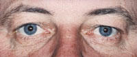
Most likely, your eyes are the first thing people notice about your appearance. They are probably the most important aspect of facial attractiveness. Unfortunately, even with a good night’s sleep, loose skin over your eyes or bags under your eyes can make you look tired or sad, or older than you really are. As we go through life, even if we have taken care of ourselves, the skin around our eyes stretches and wrinkles. Fatty deposits cause the upper lids to sag, while under the eye the tissue bulges forward and becomes discolored. In severe cases, vision can become partially blocked. CAUSES OF DROOPY EYES: Age Sun exposure Alcohol use Smoking Poor nutrition Heredity TREATMENT FOR BAGGY EYES: The most common treatment for baggy eyes is called blepharoplasty or eyelid surgery. We offer a number of finance options and affordable payment plans. It is easy to apply online and receive credit approval before you schedule an appointment.
We were all born with smooth, baby-soft skin. When we were children, the muscles under our skin contracted to display facial lines of emotion, signifying happiness or sadness. As soon as our emotions returned to normal, our expression lines disappear. But as we go through a lifetime of emotions and our skin begins to lose its elasticity, those facial lines do not go away completely, making us look older than we feel. Our skin becomes a road map showing the effects of where we have been and what we have done. CAUSES OF FACIAL WRINKLES: Age Sun exposure Alcohol use Smoking Poor nutrition Heredity TREATMENT FOR FACIAL WRINKLES: There are several cosmetic procedures that can reduce facial wrinkles. We offer a number of finance options and affordable payment plans. It is easy to apply online and receive credit approval before you schedule an appointment.
EYE CONDITIONS
Astigmatism, unlike normal vision, occurs when the cornea is shaped like a football (more curved in one direction than the other) and often occurs in combination with myopia (nearsightedness) and hyperopia (farsightedness). This causes light to focus in more than one point on the retina, resulting in blurry and distorted vision. SYMPTOMS OF ASTIGMATISM: Blurry, distorted vision at all distances CAUSES OF ASTIGMATISM: Heredity Lid swellings such as chalazia Corneal scars Keratoconus DIAGNOSING ASTIGMATISM: Your eye doctor can conduct a refractive evaluation to determine whether your eyes focus light rays exactly on the retina at distance and near. A visual acuity test will determine your ability to see sharply and clearly at all distances. Your eye doctor will also check your eye coordination and muscle control, as well as your eyes’ ability to change focus. All of these are important factors in how your eyes see. TREATMENT OF ASTIGMATISM: Glasses and contact lenses are used by many for the temporary treatment of astigmatism. However, there are a number of vision correction procedures that can surgically reduce or eliminate astigmatism. Other types of refractive errors include: nearsightedness, farsightedness and presbyopia.
Blepharitis means inflammation of the eyelids. Some doctors call it granulated eyelids. Almost everyone gets blepharitis at some time in his or her life. Some people get it repeatedly. Fortunately, blepharitis is relatively easy to treat. TYPES OF BLEPHARITIS: Staphylococcus blepharitis is caused by a germ called Staphylococci, commonly known as “staph.” It often begins in childhood and continues throughout adulthood. This form of the condition results in collar scales on lashes, crusting, and chronic redness at the lid margin. Dilated blood vessels, loss of lashes, sties, and chalazia (nodules on the eyelids) also occur. Seborrhea blepharitis is the most common and least severe form of this condition. It is not an infection but is caused by improper function of the oil glands, which causes greasy, waxy scales to accumulate along the eyelid margins. Seborrhea may be a part of an overall skin disorder that affects other areas. Hormones, nutrition, general physical condition and stress are factors in seborrhea. Ulcerative blepharitis is a less common but more severe condition that may be characterized by matted, hard crusts around the eyelashes, which, when removed, leave small sores that may bleed or ooze. There may also be a loss of eyelashes, distortion of the front edges of the eyelids and chronic tearing. In severe cases, the cornea, the transparent covering of the front of the eyeball, may also become inflamed. SYMPTOMS OF BLEPHARITIS: Itchy, burning, watery eyes Sore eyes Sticky discharge that causes the eyelashes to stick together Redness of the eyelid edges Frequent sty formation Tiny pimples on the eyelid edges Scaly skin flakes along the eyelid margins Gritty sensation leading to irritated eyes and light sensitivity Blurred vision CAUSES OF BLEPHARITIS: Poor eyelid hygiene Excess oil produced by the glands in the eyelid Bacterial infection (often staphylococcal) Allergic reaction TREATING BLEPHARITIS: In addition to eliminating redness and soreness, treatment can prevent potential infection and scarring of the cornea. You doctor will perform a complete eye examination to determine the most effective treatment. Cleaning Usually, blepharitis can be controlled by careful, daily cleaning of the eyelashes. You can do this by moistening a clean washcloth with tap water as warm as you can stand without burning. Hold the washcloth against the eyelids until it cools, then rewarm and repeat for five to ten minutes. After soaking, scrub each eyelid gently for one minute using a clean washcloth wrapped around your index finger and moistened with warm tap water. Cotton-tipped applicators, like Q-tips, are also useful to remove accumulated material from the eyelashes. To remove excessive amounts of material from your eyelids, use a few drops of a non-irritating shampoo, such as baby shampoo, mixed in lukewarm water. Being careful to avoid getting shampoo in your eye, scrub back and forth along the eyelashes of all eyelids, and then rinse with plain tap water. Once the redness and soreness are under control, this cleaning may be decreased from daily to twice weekly. However, if the symptoms return, return to daily cleansing immediately. Medication In some cases, your doctor may prescribe eye drops or ointment to be used along with the daily cleansing regimen. For ointments, use a clean fingertip to rub a small amount into the eyelashes. Be careful to follow recommended dosages; excess medication will cause temporary blurring of vision. And with any medication, there is a small possibility of allergy or other reaction. If you think this is happening, stop the medication and contact your doctor immediately. For certain types of blepharitis, medications taken by mouth are helpful. Most of these medications are antibiotics that also improve or alter the oil composition of the eyelid oil glands. When taken properly, they are safe. However, side effects may occur in some individuals, including skin rash, slight nausea and increased sensitivity to sun. Although medications may help control the symptoms of blepharitis, they alone are not sufficient; keeping the eyelids clean is essential. If you think you may have blepharitis, your eye doctor can determine the cause and recommend the right combination of treatment specifically for you.
The cornea is the clear, outer window of the eye. A corneal abrasion is simply a scratch in the epithelium (skin), or the thin, outer layer of the cornea. Abrasions usually heal in a short time period, sometimes within hours. Deeper or larger scratches may take up to a week. The cornea has a tremendous number of nerve endings, which makes any damage to the cornea very painful. SYMPTOMS OF CORNEAL ABRASIONS: History of a recent eye trauma Watery eyes Acute pain Sensitivity to light Blurry vision The feeling that there’s something in your eye Twitching eyelid CAUSES OF CORNEAL ABRASIONS: Foreign body in the eye Contact lenses Chemicals Blow to the eye Scratched eye (fingernails, hairbrushes, tree branches, etc). DIAGNOSING CORNEAL ABRASIONS: Your eye doctor can identify corneal abrasions by examining your eyes with magnifying instruments. Your doctor will check your eye, including under your eyelid, to make sure there are no foreign materials present. Depending on the initial exam, fluorescein dye may be used to help locate and identify the corneal abrasions. A test called the Seidel test (painting the wound with dye and observing for leakage) may be performed to uncover possible deeper injuries. TREATING CORNEAL ABRASIONS: Your doctor may apply a topical anesthesia to help relieve the pain. Usually, a tight patch will be placed over the eye and if the abrasion is small, the epithelium should heal overnight. If the abrasion is large, it may take a few days and your doctor may prescribe antibiotics to help prevent infections. It is important that you do not rub your eye, especially during the healing process.
he name “dry eye” can be a little confusing since one of the most common symptoms is excessive watering! It makes more sense, though, when you learn that the eye makes two different types of tears. The first type, called lubricating tears, is produced slowly and steadily throughout the day. Lubricating tears contain a precise balance of mucous, water, oil, nutrient proteins, and antibodies that nourish and protect the front surface of the eye. The second type of tear, called a reflex tear, does not have much lubricating value. Reflex tears serve as a kind of emergency response to flood the eye when it is suddenly irritated or injured. Reflex tears might occur when you get something in your eye, when you’re cutting onions, when you’re around smoke, or when you accidentally scratch your eye. The reflex tears gush out in such large quantities that the tear drainage system can’t handle them all and they spill out onto your cheek. Still another cause of reflex tearing is irritation of the eye from lack of lubricating tears. If your eye is not producing enough lubricating tears, you have dry eye. SYMPTOMS OF DRY EYE: Watery eyes The feeling that there’s sand in your eyes Eyes that itch and burn Vision that becomes blurred after periods of reading, watching TV, or using a computer Red, irritated eyes that produce a mucus discharge CAUSES OF DRY EYE: Age: As we get older, glands in the eyelid produce less oil. Oil keeps tears from evaporating off the eye. Decreased oil production allows tears to evaporate too quickly, leaving the eye too dry. Diseases including diabetes, Sjogren’s and Parkinson’s Hormonal changes, especially after menopause Prescription medications: These include some high blood pressure medications, antihistamines, diuretics, antidepressants, anti-anxiety pills, sleeping pills and pain medications. Over-the-counter medications including some cold and allergy products, motion sickness remedies, and sleep aids can also cause dry eye. Hot dry or windy conditions: High altitude, air-conditioning and smoke can also cause dry eye. Reading, using a computer or watching TV Contact lenses Eye surgery: Some types of eye surgery, including iLASIK can aggravate dry eye. Inflammation: Recent research suggests that dry eye may be caused by inflammation due to an imbalance of “good” fats and “bad” fats. DIAGNOSING DRY EYE: Your eye doctor can check for dry eye by examining your eyes with magnifying instruments, measuring your rate of tear production and checking the amount of time it takes for tears to evaporate between blinks. The doctor can also check for pinpoint scratches on the front surface of the eye caused by dryness using special, colored eyedrops call fluorescein or Rose Bengal. TREATMENTS FOR DRY EYE: The most common treatment is use of artificial teardrops that help make up for the lack of natural lubricating tears. Artificial tear products come in liquid form, longer lasting gelform and long-lasting ointment form, which is most often recommended for nighttime use. Many different brands of artificial tears are available over-the-counter. Some contain preservatives and some do not. Unpreserved tears may be recommended for people whose eyes are sensitive to preservatives. Artificial tears can generally be used as often as needed, from a few times per day to every few minutes. You should follow the regimen your doctor recommends. When infection, inflammation of the eyelids or clogged oil glands contribute to dry eye, special lid cleaning techniques or antibiotics may be recommended. It may also help to avoid hot, dry or windy environments or to humidify the air in your home or office. Restasis is an exciting new treatment for Dry Eye Disease. Restasis drops help the eyes produce more tears by reducing inflammation, which is often a cause of dry eye. Unlike artificial tears, Restasis is the first drug proven to effectively treat a cause of Dry Eye Disease rather than only temporarily alleviate symptoms. Punctal occlusion is a medical treatment for dry eye that may enable your eyes to make better and longer use of the few lubricating tears they do produce.
Macular degeneration is a disease of the macula, an area of the retina at the back of the eye that is responsible for fine detail vision. Vision loss usually occurs gradually and typically affects both eyes at different rates. Even with a loss of central vision, however, color vision and peripheral vision may remain clear. SYMPTOMS OF MACULAR DEGENERATION: Early macular degeneration may cause little, if any noticeable change in vision Difficulty reading without extra light and magnification Seeing objects as distorted or blurred, or abnormal in shape, size or color The perception that objects “jump” when you try to look right at them Difficulty seeing to read or drive Inability to see details Blind spot in center of vision There are two forms of age-related macular degeneration, wet and dry. WET MACULAR DEGENERATION Wet macular degeneration occurs when abnormal or leaking blood vessels grow underneath the retina in the area of the macula. These changes can lead to distorted or blurred vision and, in some cases, a rapid and severe loss of straight ahead vision. DRY MACULAR DEGENERATION The vast majority of cases of macular degeneration are the dry type, in which there is thinning or deterioration of the tissues of the macula or the formation of abnormal yellow deposits called drusen. Progression of dry macular degeneration occurs very slowly and does not always affect both eyes equally. CAUSES OF OR CONTRIBUTING FACTORS TO MACULAR DEGENERATION: The root causes of macular degeneration are still unknown. Women are at a slightly higher risk than men. Caucasians are more likely to develop macular degeneration than African Americans. Age: Macular degeneration is the leading cause of decreased vision in people over 65 years of age. Heredity: Macular degeneration appears to be hereditary in some families but not in others Long-term sun exposure Smoking High blood pressure High cholesterol Hypertension Nutritional deficiencies Diabetes Head injury Infection DIAGNOSING MACULAR DEGENERATION: Your eye doctor can identify changes of the macula by looking into your eyes with various instruments. A chart known as an Amsler Grid can be used to pick up subtle changes in vision. Please go to Patient Forms to download the Amsler Grid test and receive instructions on how to test your vision at home. Angiography is the most widely used macular degeneration diagnostic test. During the test, a harmless orange-red dye called Fluorescein will be injected into a vein in the arm. The dye travels through the body to the blood vessels in the retina. A special camera takes multiple photographs. The pictures are then analyzed to identify damage to the lining of the retina or atypical new blood vessels. The formation of new blood vessels from blood vessels in and under the macula is often the first physical sign that macular degeneration may develop. Optical Coherence Tomography (OCT) uses light waves to create a contour map of the retina and can show areas of thickening or fluid accumulation. TREATMENT FOR MACULAR DEGENERATION: In the early stages of macular degeneration, regular eye check-ups, attention to diet, in-home monitoring of vision and possibly nutritional supplements may be all that is recommended. DIET AND NUTRITIONAL SUPPLEMENTS There has been active research on the use of vitamins and nutritional supplements called antioxidants to try to prevent or slow macular degeneration. Antioxidants are thought to protect against the damaging effects of oxygen-charged molecules called free radicals. A potentially important group of antioxidants are called carotenoids. These are the pigments that give fruits and vegetables their color. Two carotenoids that occur naturally in the macula are lutein and zeaxanthin. Some research studies suggest that people who have diets high in lutein and zeaxanthin may have a lower risk of developing macular degeneration. Kale, raw spinach, and collard greens are vegetables with the highest amount of lutein and zeaxanthin. You can also buy nutritional supplements that are high in these and other antioxidants. LOW VISION AIDS Unfortunately, the vast majority of cases of wet macular degeneration and virtually all cases of dry macular degeneration are not treatable. In these cases, low vision aids may help make it easier to live with the decreased vision of macular degeneration. Low vision aids range from hand-held magnifying glasses to sophisticated systems that use video cameras to enlarge a printed page. Lifestyle aids such as large print books, tape-recorded books or magazines, large print playing cards, talking clocks and scales and many other devices are available. INJECTION LUCENTIS and Macugen are new treatments for the wet form of age-related macular degeneration. These injections block abnormal blood vessel growth and leakage.
We were all born with smooth, baby-soft skin. When we were children, the muscles under our skin contracted to display facial lines of emotion, signifying happiness or sadness. As soon as our emotions returned to normal, our expression lines disappear. But as we go through a lifetime of emotions and our skin begins to lose its elasticity, those facial lines do not go away completely, making us look older than we feel. Our skin becomes a road map showing the effects of where we have been and what we have done. CAUSES OF FACIAL WRINKLES: Age Sun exposure Alcohol use Smoking Poor nutrition Heredity TREATMENT FOR FACIAL WRINKLES: There are several cosmetic procedures that can reduce facial wrinkles. We offer a number of finance options and affordable payment plans. It is easy to apply online and receive credit approval before you schedule an appointment.
A pterygium is a fleshy triangular tissue that grows over the cornea, usually on the inner corner of the eye. Sometimes, it grows big enough that it interferes with vision. As the pterygium develops, it may alter the shape of the cornea, causing astigmatism. SYMPTOMS OF A PTERYGIUM: Tissue growing over the eye Irritation Redness Tearing CAUSES OF A PTERYGIUM: The exact cause of pterygia isn’t known, but it’s thought to be linked to: Long-term exposure to sunlight Dry, dusty conditions Age: Ptergia is typically found in adults over the age of 30. DIAGNOSING A PTERYGIUM: Chances are, you’ll notice a pterygium. Your eye doctor can also diagnose it during a routine eye exam. TREATMENT OF A PTERYGIUM: Eye drops or ointment can be used to reduce the irritation caused by a pterygium. If the pterygium grows toward the central cornea,it may need to be removed surgically. Prevention is important. It’s a good idea to protect your eyes with sunglasses if you’ll be in the sun or a dusty, dry environment.
Strabismus is a condition in which the eyes point in different directions. Types of strabismus include crossed eyes (esotropia), out-turned eyes (exotropia), or vertical misalignment (hyper or hypotropia). Turning of the eye may be constant or may show up only some of the time. Strabismus should be evaluated and treated. SYMPTOMS OF STRABISMUS Double vision Eyes that look in different directions Head tilt or turn CAUSES OF STRABISMUS Strabismus is caused by a lack of coordination between the eyes. In children, the cause of strabismus is not known, although the condition runs in families and occurs more commonly in children with neurological problems. In adults, causes of strabismus include injury to an eye muscle or the nerves controlling those muscles; head trauma; conditions such as diabetes or high blood pressure; loss of vision; an eye or brain tumor; Graves’ disease, stroke or other muscle and nerve disorders. TREATING STRABISMUS Children with strabismus must be identified and treated at a young age in order to prevent permanent vision problems. In children, when the two eyes fail to focus on the same image, the brain may learn to ignore the input from one eye. If this is allowed to continue, the eye that the brain ignores will never see well. Children do not grow out of strabismus. Treatment options depend upon the type of strabismus and may include glasses, patching one eye, prism lenses and/or surgery.
LASER VISION CORRECTION
ILASIK PROCEDURE: You’ve heard thousands of LASIK ads touting thousands of different things, but the iLASIK procedure ends all of the noise. It’s all you will need to know – outstanding technology, one brand and one easy way to better vision. YOU CAN AFFORD IT: The iLASIK procedure is more affordable than you think Hattiesburg Eye Clinic works with CareCredit to help you finance the procedure with monthly payments to fit your budget. STRAIGHT TALK ABOUT THE ILASIK PROCEDURE: A lot of people have had successful LASIK, but as many as 10 times that number are good candidates that are waiting. Some people are waiting because they don’t think they can afford it. Others are unaware of the true safety of the procedure. But most people are waiting simply because they lack the right information. The introduction of the iLASIK procedure means the wait is over and it’s simple: SAFE & PROVEN: The technology used in the iLASIK procedure is very mature (the systems used today have been in use for years) and very safe. Doctors have been performing LASIK for a decade. Approximately 17 million LASIK procedures have been performed to date, making it the most common elective vision procedure in the U.S. In fact, all branches of the U.S. military and NASA recently approved LASIK for their servicemen and women, thanks to studies using iLASIK technology. ABOUT THE PROCEDURE: The iLASIK procedure only takes minutes. It reshapes your cornea to correct the refractive imperfections in your eye (these imperfections are essentially the reason you are nearsighted, farsighted, etc.). Here’s the procedure in a nutshell: HERE’S WHAT HAPPENS: You first receive an extensive exam by an iLASIK surgeon We use computer software to make an individual blueprint of your eyes; we then use this blueprint to drive your laser correction to give you a truly customized result You’ll be given numbing drops before we begin so you won’t feel pain A laser creates a thin flap in your cornea This flap enables the surgeon to make the iLASIK correction We use a second laser to make the correction to your cornea, based on your unique vision and eye characteristics This correction is completely computer driven It’s safe, fast and virtually painless You can have great vision without glasses or contacts* Outcomes may vary. 98% of nearsighted patients participating in the Advanced CustomVue™ FDA Clinical Trials could see 20/20 or better one year after treatment. THE TECHNOLOGY BEHIND ILASIK: To correct your vision with the iLASIK procedure, a thin corneal flap needs to be created. There are two ways to make this flap: a blade or the IntraLase™ FS laser. The iLASIK procedure EXCLUSIVELY uses the IntraLase laser; it’s part of what makes iLASIK vision correction unique. Eighty-one percent of patients choose bladeless LASIK over traditional LASIK with a blade when given the choice. FREQUENTLY ASKED QUESTIONS: What is LASIK? LASIK is an acronym that stands for laser in-situ keratomileusis. It’s a surgical procedure using laser technology to lift a thin layer of cornea in such a way as to create a flap that stays connected on one side. These flaps are usually only a hundred microns thick, which is quite thin. After the laser does its job on the flap, we then move the patient to another laser that sculpts the cornea with a cool, ultraviolet light into the right shape to correct the patient’s refractive problem, be it nearsightedness, farsightedness or astigmatism. We then lay the flap down to its original position where it takes about a minute for the cornea to dry to allow the flap to remain adhered. The actual laser time for all the steps in this procedure for most patients is about a minute. What’s the difference between nearsightedness and farsightedness? The difference between nearsightedness (myopia) and farsightedness (hyperopia) depends on how light rays enter the eye. Light is mostly refracted (changes direction) at the corneal surface at the very front part of the eye. If the shape of the cornea refracts to focus the light rays to the front of the retina, a person can experience nearsightedness where they can see normally up close but their distance vision is blurry. Farsightedness occurs when the shape of the cornea refracts light to focus past the retina. In this case, patients can’t see clearly at either near or far distances. To correct these two problems we must essentially change the refraction of the light rays so that they focus at the correct location in reference to the retina. Traditionally, we can do this with glasses or contacts, and now we can do the same with refractive surgery like LASIK.
PRK (Photo-Refractive Keratectomy) was the first procedure performed using the Excimer laser. It corrects vision by reshaping the cornea. The difference between LASIK and PRK is that with LASIK a corneal flap is created and the laser is applied to the inner tissue of the cornea. With PRK, the epithelium (or outer skin of the cornea) is removed and a laser is applied to the surface of the cornea. PRK can be used to correct low to high levels of nearsightedness, farsightedness and astigmatism. To treat nearsightedness, the steep cornea is made flatter by removing tissue from the center of the cornea. This moves the point of focus from in front of the retina to directly on the retina. To treat farsightedness, the flat cornea is made steeper by removing tissue outside of the central optical zone of the cornea. This moves the point of focus from behind the retina to directly on the retina. To treat astigmatism, the cornea is made more spherical — like a basketball instead of a football. This eliminates multiple focusing points within the eye and creates one point of focus on the retina. Astigmatism can be treated at the same time as nearsightedness and farsightedness. PRK IS FOR THOSE WHO: want to reduce or eliminate their dependence on glasses or contacts are over 18 years of age have had a stable eye prescription for at least one year have no health issues affecting their eyes have corneas too thin for LASIK have large pupils WHAT TO EXPECT ON SURGERY DAY: You will arrive at the laser center about an hour prior to your procedure. Once you have been checked in you may be offered a sedative to help you relax. You will then be prepared for surgery. The area around your eyes will be cleaned and a sterile drape may be applied around your eye. Anesthetic eye drops will be used to numb your eyes; no injections or needles will be used. When your eye is completely numb, an eyelid holder will be placed between your eyelids to keep you from blinking during the procedure. Next, your doctor will remove the epithelium, a thin layer of protective skin that covers the cornea. You will be asked to look directly at a target light while the laser reshapes your cornea. The laser will be programmed with the information gathered in your pre-operative exam. The laser treatment will be completed in less than a minute or two, depending on the amount of correction needed. Following your procedure, your eye(s) will be examined with a slit lamp microscope. Your eyes may be shielded for protection. Your vision may be blurry or hazy for one to five days. You will experience some discomfort as the epithelium heals and covers the treated area. Eye drops, pain medication and possibly a protective contact lens can be used to minimize this discomfort. Most patients resume normal activities within one to three days. Vision can fluctuate for up to six months. REALISTIC EXPECTATIONS: The decision to have PRK is an important one that only you can make. The goal of any refractive surgical procedure is to reduce your dependence on corrective lenses. However, we cannot guarantee you will have the results you desire. The vast majority of our patients are extremely happy with their vision after PRK and can do most activities without dependence on corrective lenses. PRK is a safe, effective and permanent procedure, but like any surgical procedure, it does have some risks. After PRK, almost everyone experiences some visual side effects.These visual side effects are usually mild and temporary and have a tendency to diminish over time. But there is a slight chance that some of these side effects won’t go away completely, including light sensitivity, glare and halos. Serious complications to PRK are extremely rare. Since everyone heals somewhat differently, some patients may over react to the procedure and some may under react resulting in overcorrections and undercorrections. Once the eye has stablized (3 to 6 months) you and your doctor can discuss whether a re-treatment could help fine tune your vision if you are over or under corrected. After a thorough eye exam, you and your doctor will determine if PRK is an option for you. If you are a good candidate, you will be given additional information about the procedure that will allow you to make an informed decision about whether to proceed.Be sure you have all your questions answered to your satisfaction. ALTERNATIVES TO PRK PRK is not the only surgical procedure designed to correct nearsightedness, farsightedness and astigmatism. To learn about other procedures go to the surgical and laser vision correction procedures section of our Web Site. If you would like to learn more about vision correction procedures from sources other than our practice, we encourage you to link to a number of Web sites we feel provide factual and up-to-date information. You may also choose to make an appointment or request additional information to learn more about this exciting procedure.
As we age, the lens inside our eye loses its ability to focus both near and far. Most vision correction procedures attempt to change the focusing power of the cornea. A refractive lens exchange, on the other hand, corrects nearsightedness or farsightedness by replacing the eye’s natural lens, which has the wrong power, with an artificial intra-ocular lens (IOL) implant that has the correct power for the eye. A refractive lens exchange is a surgical procedure that uses the same successful techniques of modern cataract surgery. These surgical techniques have evolved and improved dramatically over the last 20 years. Cataract surgery is now the most common surgical procedure performed in medicine today. The main difference between standard cataract surgery and a refractive lens exchange is that cataract surgery is primarily performed to remove a patient’s cataract that is obstructing and clouding their vision, while a refractive lens exchange is performed to reduce a person’s dependence on glasses or contact lenses. A refractive lens exchange can be combined with other procedures that treat astigmatism such as LRIs and AK. A REFRACTIVE LENS EXCHANGE IS FOR THOSE WHO: want to reduce or eliminate their dependence on glasses or contacts may not be a good candidate for laser vision correction are 18+ years of age have no health issues affecting their eyes Cataracts are a part of the normal aging process and if a person lives long enough chances are they will develop cataracts. People who have refractive lensectomy now, will not have to have cataract surgery in the future. WHAT TO EXPECT ON SURGERY DAY: The refractive lens exchange procedure is performed on an outpatient basis. Only one eye will be treated at a time. After the eye is completely numbed with topical or local anesthesia, the eye’s natural lens will be gently vacuumed out through a tiny incision, about one eighth of an inch wide. Next, the new, intra-ocular lens will be folded and inserted through the same micro-incision. It will then be unfolded and placed into the “capsular bag” that originally surrounded the natural lens. This incision is “self-sealing” and usually requires no stitches. It remains tightly closed by the natural outward pressure within the eye. This type of incision heals fast and provides a much more comfortable recuperation. ACRYSOF® RESTOR® IOL For most RLE patients, life without glasses or bifocals is something they either experienced before age 40, or they just dreamed about for most of their lives. But today, the AcrySof® ReSTOR® IOL is providing our patients with the ability to see clearly at all distances without dependence on bifocals or reading glasses. Each of these presbyopia correcting IOLs is unique in its optical design and visual advantages. Which one is best for you depends on the unique characteristics of your eye as well as your lifestyle needs. After a thorough eye examination we will make a recommendation as to which of these revolutionary IOLs would be most advantageous for you. If your eye has pre-existing astigmatism, your surgeon may elect to make micro-incisions in the cornea to reduce your astigmatism. These are called LRIs or limbal relaxing incisions. You will go home soon after the surgery and relax for the rest of the day. Everyone heals somewhat differently but many patients report improvement in their vision almost immediately after the procedure. Most patients return to their normal activities within a day or two. REALISTIC EXPECTATIONS: The decision to have a refractive lens exchange is an important one that only you can make. The goal of a refractive lens exchange is to reduce or eliminate your dependence on glasses or contact lenses. However, we cannot guarantee you will have the results you desire. Serious complications with refractive lens exchange are extremely rare. It is a safe, effective and permanent procedure, but like any surgical procedure, it does have some risks. Going to an eye specialist experienced with the procedure can significantly minimize the risks involved with refractive lensectomy. After a thorough eye exam, you and your doctor will determine if refractive lensectomy is an option for you. If you are a good candidate, you will be given additional information about the procedure that will allow you to make an informed decision about whether to proceed. Be sure you have all your questions answered to your satisfaction. ALTERNATIVES TO REFRACTIVE LENSECTOMY A refractive lens exchange is not the only surgical procedure designed to correct nearsightedness. To learn about other procedures go to the surgical and laser vision correction procedures section of our Web Site. If you would like to learn more about vision correction procedures from sources other than our practice, we encourage you to link to a number of Web sites we feel provide factual and up-to-date information.
LASEK (Laser Epithelial Keratomileusis) is a relatively new laser vision procedure that combines elements of PRK and LASIK, and may offer some advantages over both. Instead of removing the epithelium (the thin layer of protective skin that covers the cornea) like in PRK, a flap of surface epithelium is loosened with a diluted alcohol solution and moved aside. The surface under the epithelium is then treated with the laser and the epithelial flap is returned to its original position. Using the epithelium flap as a natural protective bandage with LASEK, as opposed to completely removing the epithelium as with PRK, may improve healing and the incidence of postoperative haze. The margin of safety with LASEK may be greater than that with LASIK as the need for a microkeratome is eliminated, so there’s no risk of flap complications. LASEK removes tissue within the cornea to treat low to high levels of nearsightedness, farsightedness and astigmatism. To treat nearsightedness, the steep cornea is made flatter by removing tissue from the center of the cornea. This moves the point of focus from in front of the retina to directly on the retina. To treat farsightedness, the flat cornea is made steeper by removing tissue outside of the central optical zone of the cornea. This moves the point of focus from behind the retina to directly on the retina. To treat astigmatism, the cornea is made more spherical — like a basketball instead of a football. This eliminates multiple focusing points within the eye and creates one point of focus on the retina. Astigmatism can be treated at the same time as nearsightedness and farsightedness. LASEK IS FOR THOSE WHO: want to reduce or eliminate their dependence on glasses or contacts are over 18 years of age have had a stable eye prescription for at least one year have no health issues affecting their eyes have wide pupils have corneas too thin for LASIK who do not want LASIK because it requires a microkeratome WHAT TO EXPECT ON SURGERY DAY: You will arrive at the laser center about an hour prior to your procedure. Once you have been checked-in and settled comfortably, you will be prepared for surgery. The area around your eyes will be cleaned and a sterile drape will be applied. You may be given a sedative to help you relax. Eye drops will be used to numb your eyes; no injections or needles are used. When your eye is completely numb, an eyelid holder will be placed between your eyelids to keep you from blinking. Next, a specially designed instrument will be placed on your eye around the area that will have the laser applied. Then a diluted alcohol solution will be placed in the instrument, which loosens the epithelium from the surface of the cornea. The epithelium is then pushed to the side. You will be asked to look directly at a target light while the laser reshapes your cornea. The laser will be programmed with the information gathered in your pre-operative exam. The laser treatment will be completed in less than a minute or two, depending on the amount of correction needed. To finish the procedure, your doctor will smooth the flap back into place. A temporary bandage contact lens will be used to keep the epithelium in place and reduce post-operative discomfort. Following your procedure, your eye(s) may be shielded for protection. Your vision will probably be a little blurry at first, so someone will need to drive you home. You should relax for the rest of the day. You may experience mild to moderate discomfort for two or three days. You will be given instructions on how to manage this discomfort before you leave. Most patients resume normal activities within two to three days. Vision can fluctuate for up to six months. REALISTIC EXPECTATIONS: The decision to have LASEK is an important one that only you can make. The goal of any refractive surgical procedure is to reduce your dependence on corrective lenses. However, we cannot guarantee you will have the results you desire. The vast majority of our patients are extremely happy with their vision after LASEK and can do most activities without dependence on corrective lenses. Serious complications with LASEK are extremely rare. LASEK is a safe, effective and permanent procedure, but like any surgical procedure, it does have some risks. Many of the risks and complications associated with this procedure can be reduced or eliminated through careful patient selection and thorough pre-operative testing using the latest diagnostic technology. After LASEK, you may experience some visual side effects. These visual side effects are usually mild and most often diminish over a few days to a few weeks. But there is a slight chance that some of these side effects won’t go away completely, such as feelings of dryness, glare and halos.
PEDIATRIC SERVICES
While the preceding visit schedule is a good guideline, you don’t have to wait to see us if you notice anything out of the ordinary with their vision. Keep an eye out, then, for any of the following abnormal signs: Your child squints, rubs their eyes or is sensitive to light Has a sudden drop in grades or can’t copy from the board Avoids reading or uses their finger to follow as they read Seems confused over left or right or has other poor directional skills Demonstrates poor hand-eye coordination Sits too close to a viewing screen Tilts their head to see better Experiences chronic headaches or excessive eye tearing These all could indicate a potential vision problem. If you see any of them, don’t hesitate to call us at (601) 268-5910 to request an appointment. GOOD VISION IS CRUCIAL TO LEARNING AND EARLY SOCIAL DEVELOPMENT Especially for school-age children, vision is important for learning. It’s estimated up to 80% of what we learn comes through seeing and reading. It’s also estimated that one in four school-age children have some form of vision problem that could impede learning – and one in ten need glasses to correct it. To make matters worse, children don’t always notice when they have a vision problem and may not complain; problems like Amblyopia (lazy eye) or nearsightedness may not have apparent symptoms. Regular, comprehensive vision exams, especially before your child enters school, may be the only way to diagnose such problems early. An unrecognized, untreated vision problem could also have an adverse effect on more than their ability to learn. Because of poor vision they may not be able to participate fully in sports or other activities, which could limit their social and emotional development. CALL US FOR MORE INFORMATION OR TO MAKE AN APPOINTMENT Hattiesburg Eye Clinic’s Pediatric Vision Team can make a difference in your child’s eye and vision health. To learn more or request an appointment, call (601) 268-5910 and ask about our pediatric vision exams and services.
VARIOUS EYE PROBLEMS AN EYE EXAM CAN REVEAL There are several possible conditions we look for when we examine a child’s eyes. The following are a few such conditions a comprehensive visual exam can identify, and which call for prompt, early treatment to minimize visual impairment, Common Refractive Problems. The parts of the eye work together to transmit received light as a focused image in the brain. When they don’t, a person experiences out-of-focus vision, blurring of objects at a distance (nearsightedness), up close (farsightedness), or as a result of distortions in the lens or cornea (astigmatism). Strabismus. Children with this condition are unable to align both eyes at the same time as they look at a single point. The misalignment is often called wandering eye or “cross eyes” because of their appearance. Amblyopia. Also known as “lazy eye,” Amblyopia causes one of a child’s eyes to have poorer vision than the other. As their visual function matures, the brain may begin to “ignore” the vision from the faulty eye. Ptosis. This occurs when the upper eyelid droops down abnormally over the eye. While in some cases the droop may be slight, it’s possible for the lid to completely cover the eye pupil and interfere with vision. Tear duct obstruction. For any number of reasons when a tear duct becomes blocked, tears can’t drain properly through the nasal passages. The obstruction can cause excessive tearing as the fluid overflows instead out of the eye. Blepharitis. In this common condition, an eyelid can become reddened, swollen (inflamed) and itchy and lead to dry eye. Causes for Blepharitis include bacterial buildup, blockage of the eyelid’s oil glands or allergies. Ocular allergies. Allergies occur when a person’s immune system overreacts to a foreign substance. In the eye, this can cause irritation of the conjunctiva, a mucous membrane on the front of the eyes and the inside of the eyelids. WHY VISION EXAMS BEFORE AGE ONE? Good vision includes more than an ability to see clearly. Your child may be able to see “20/20,” yet still have issues with normal eye movement or a lack of coordination in focusing that could affect depth perception. A comprehensive vision examination will check for acuity (ability to see clearly), binocularity (both eyes working together to perceive depth), eye movement and focusing ability, and overall eye health. By beginning with an initial exam in their first year and then at regular intervals from then on, we can track their vision development, diagnose problems as they arise and initiate treatment. As in many other aspects of health, the earlier we can recognize a problem and begin treating it, the better the chances of a successful outcome. CALL US FOR MORE INFORMATION OR TO MAKE AN APPOINTMENT Hattiesburg Eye Clinic’s Pediatric Vision Team can make a difference in your child’s eye and vision health. To learn more or request an appointment, call (601) 268-5910 and ask about our pediatric vision exams and services.
Reviews
Brand Certified Facts from Hattiesburg Eye Clinic
- Address
- Categories
- Geo coordinates
- Legal business name
- Hours of operation
- Phone number
- Official website